


How to maintain a microscope

Microscope maintenance is strongly advised in order to prolong its life and accuracy. It is always recommended that school microscopes be professionally serviced. However, it is prudent for the microscopes to be cleaned regularly by school Science Technicians too.
This guide explains how this can be done by the Technician. Please note that the method will vary depending on the microscope age and design.
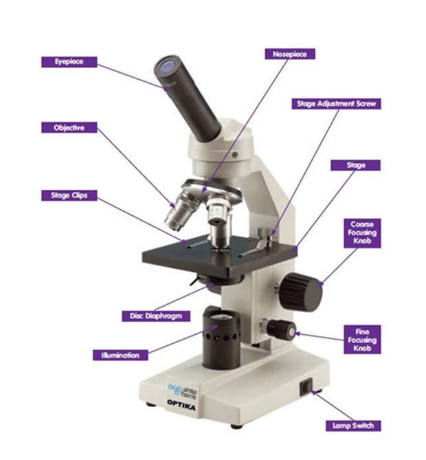
Parts of a microscope
Before cleaning and servicing the microscope, here is a refresher on the parts of a microscope
Cleaning and servicing a microscope
Removal of the optics
- Lay out a piece of paper next to the microscope where you are planning to work
- Remove the eyepiece from the instrument as a unit and lay this on the paper
Some eyepieces are locked into the microscope tube with grub screws; an instrument screwdriver or Allen key will be required to loosen them. Some eyepieces are just a push fit in the body tube and can be lifted out; others need to be unscrewed bodily.
- Ensure that the whole eyepiece (two lenses) has been removed; the tube should be clear for at least the depth of a finger
- Remove the objectives and lay these on the paper
Most objectives unscrew; use two hands to ensure the objective does not drop. Some objectives have a locking grub screw which must be loosened first. Some narrower objectives (without milled collars) can be gripped more easily by sliding a short piece of rubber tubing over them.
Cleaning the optics
Optical glass is easily scratched and its surface can be damaged by repeated immersion in water. The mountants holding the component lenses in objectives and condensers can be attacked by alcohol and other organic solvents. If possible, lenses should be cleaned by:
- Puffing air across them from hand bellows (not the mouth!), dusting with an artists’ soft paint brush or wiping with a lens tissue moistened with distilled water. Only use organic solvents to remove contamination where water is unsuccessful. Wipe the surface gently with a lens tissue moistened with ethanol or dimethylbenzene (xylene)
Accessible optical surfaces can be given a final polish with a microfibre spectacle-cleaning cloth or lens cleaning tissues.
Cleaning the mechanical parts
- If the grease on the fine-focus mechanism is clean and soft, leave it alone. If the grease is dirty or lumpy, remove dirt and lumps with a rag moistened with light oil rather than a grease solvent. A cocktail stick may help to push the rag into odd corners. Do not try to remove all the grease; if it is really bad, expert help is required
- Remove dirt or grease from any racks and pinions of the coarse-focusing mechanism with a grease solvent. Do not saturate with solvent and do not use acetone (propanone), carbon tetrachloride or petrol as all of these are hazardous or now illegal to use in schools. A small, stiff brush may be useful here (not the paint brush used for lenses!)
- Clean the nosepiece with a rag moistened with grease solvent
- Clean the stage, foot, limb, stand, body tube and other non-optical external parts with a rag dampened with soapy water or with a spray-foam cleaner. Difficult stains or corrosion may be tackled with appropriate cleaners but do not over-wet the parts!
Checking the microscope
After cleaning and re-assembling it is now advisable to check that the system is clean and working well.
Select a microscope slide with a well-stained section several millimetres across. Put it on the stage. Do the stage clips hold it securely but not so tightly that they prevent movement? Adjust if necessary by bending the clips slightly with a pair of pliers.
Select the lowest-power objective and the eyepiece with the widest field of view (so that as much as possible of the object can be seen at once). Illuminate the slide and focus on it using the following technique.
- Looking from the side, lower the stage until it is as low as it can go
- Looking through the microscope, raise the stage until the image is roughly in focus using the coarse focus. If this cannot be achieved, repeat the first step and/or check the reassembly of the stops and the fine-focus mechanism
- Looking from the side, check that the fine-focus mechanism produces a total movement of between one and two millimetres. There is often an indication consisting of a mark which moves between two fixed ones
- If an adjustable stop is fitted to prevent contact between objectives and slide, set it to prevent further movement of the coarse focusing when the fine focus is mid-range and the image is in focus. Do not tighten any locking nut yet
Check the eyepiece(s) for cleanliness: rotate it/them while looking at the focused image. If dark shadows or specks rotate too, the eyepiece needs further attention.
Check the objectives by selecting each one in turn. If a sharp image cannot be obtained, the bottom lens of the objective is probably still dirty. Try re-cleaning it.
Also check the setting of the focusing stop; it should be possible to focus the image using only the fine control with the coarse control against the stop. If adjustment is necessary, set the stop for the highest-power objective, taking great care not to crash the objective into the slide. Tighten any locking nut or wheel.
Then check that the microscope is still par-focal, i.e. as you switch from one objective to another, they are all more or less in focus. If parfocality has been lost, check that the objectives are the correct ones for the model of microscope being serviced.
Finally, check the condenser. Focus the microscope on a slide using the low-power objective with the condenser as high as possible and the diaphragm open as wide as possible. Remove the eyepiece and check that the disc of light seen at the bottom of the tube fills the view. If this is not possible, check the alignment and cleanliness of the condenser.
Replace the eyepiece and lower the condenser slightly to see if it improves the image. Reducing the size of the diaphragm aperture may further improve the contrast. This setting will be different for each objective.
Microscope maintenance summary
Microscopes don’t like fingertips and humidity. The prisms and lenses have anti-fungus coating which needs protecting. It is better to cover the microscope when not in use.
General cleaning: Remove dust from the eyepieces using lens tissues, paint brush or air.
Remove dust from the microscope body using a duster and always cover when not in use.
Learn all about maintaining a microscope
The Philip Harris specialist team are here to help with any technical questions you have with maintaining a microscope, or any other support you need. Call them on 0345 120 4521 or e-mail techsupport@philipharris.co.uk.
Check out our Twitter and Facebook for more hints and tips for school science laboratory technicians.


